After the Dissection
A porter carrying cadavers through Florence could find nowhere to stop with his basket. His deliveries supplied the workshop where Clemente Susini and his collaborators turned short-lived dissections into wax anatomy for students, emperors and museums.
What's the deal?
Students could study exposed organs, vessels and nerves long after the original dissection had decayed. In eighteenth-century Florence, Susini and his collaborators reproduced carefully prepared anatomy in coloured wax. Teaching collections could be copied and sent to other cities. They gave learners lasting three-dimensional views inside the body, while preserving the anatomists' choices and sometimes their mistakes.
On 19 October 1792, Giacinto Guidetti complained about the consequences of carrying dead bodies through Florence. His work for the royal museum included fetching cadavers from the hospital of Santa Maria Nuova. In rain or snow he could find nowhere to stop with his basket. People treated him badly in the coffee houses he had previously frequented.
Guidetti was a porter and cleaner, far removed from the patrons whose names adorned the museum. Yet the anatomical waxes being made there depended on his deliveries. A surgeon needed fresh material to dissect; a modeller needed the surgeon’s preparation. The serene faces and carefully coloured organs in the glass cases began with this less presentable traffic between hospital and workshop.
A hand could be opened once and studied for generations.
In the collection made for Cagliari by the Florentine modeller Clemente Susini and the anatomist Francesco Antonio Boi, two left hands lie palm upwards. One retains its skin. The other exposes muscles and nerves. Enlarged preparations of fingers make smaller structures visible. The set gives a learner several views of a hand without requiring the intact one to be destroyed.
A museum with a workshop inside
The undertaking had begun in the early 1770s under Felice Fontana, a natural philosopher who became director of the grand duke’s museum of physics and natural history. His patron was Peter Leopold, ruler of Tuscany and brother of the Habsburg emperor Joseph II. Fontana wanted an extensive artificial anatomy: a collection in which the human body could be examined in three dimensions, beyond the short working life of a dissection.
Florence already had a connection to anatomical waxwork through Giuseppe Galletti, a surgeon who had seen teaching models in Bologna. He engaged the sculptor Giuseppe Ferrini to make obstetrical and anatomical preparations. Fontana brought Ferrini into the museum’s workshop. In 1773 he recruited a second modeller and assistant to the dissector, a nineteen-year-old Florentine called Susini.
Susini arrived with an artist’s training. He had studied at Florence’s academy, won prizes for sculpture and worked in the studio of Pompilio Ticciati. His experience included glass painting, bronze sculpture and copper engraving. Wax required a different handling of colour and surface, but he was accustomed to making an object resemble something else.
When the museum opened to the public in February 1775, its anatomical collection already occupied six rooms. It became known as La Specola after the observatory installed above it. The intended audience extended beyond future doctors. Visitors could study the human body in the same institution that arranged the other productions of nature for their inspection.
This helps explain why some of the wax bodies look as though they belong to sculpture. A reclining woman rests on a pillow; a male figure raises an arm or adopts a pose familiar from monumental art. The positions could expose anatomy while giving the figure a recognisable human form. The female model later called the Venus allowed sections to be lifted away, revealing deeper organs and a fetus. Removing a layer did not ruin it: it could be put back.
What the anatomist had to see
The figures were the work of a team. Antonio Matteucci, one of the museum’s early dissectors, set out the responsibilities plainly. He had to remove the membranes and connective material that obscured the parts, using an anatomist’s judgement to make the structures clear enough to imitate. If an anatomical error resulted, he said, responsibility belonged to him. Ferrini was to follow his instructions on anatomical accuracy.
A printed atlas might suggest what to seek, but a body still had to yield the preparation. Ernst Friedrich Wichelhausen, describing Florentine manufacture in 1798, explained that engravings guided the dissector rather than replacing the cadaver. Several bodies might be needed to obtain a sufficiently complete demonstration. A finished wax could combine observations from different preparations, suppressing an awkward peculiarity here and making a hidden junction legible there.
The supplies came from institutions whose records usually tell us much more about quantities than about individuals. Santa Maria Nuova supplied adult cadavers; the Innocenti foundling institution supplied children’s bodies. The surviving administrative trail does not allow the people behind most models to be named. A lifelike face cannot by itself establish that the wax records the features of the person whose internal anatomy was dissected.
The workshop’s equipment connected this anatomical labour to ordinary craft operations. Purchase records list copper vessels for melting wax, iron wire of different thicknesses, modelling tools and marble slabs on which wax could be rolled thin. Slates allowed notes and drawings to be made during dissections. Containers held dyes and other ingredients; baskets carried the anatomical material.
How a dissection became wax
A plaster mould could be taken from the dissected preparation, or from an intermediate model in clay or rough wax. Inside its negative surface, the workers laid coloured, translucent wax in successive layers. Separate cast sections could be joined by warming their edges. Filling material and metal supports helped larger models keep their shape.
The cast still needed finishing. Vessels and nerves too delicate for the main mould were added separately; the workshop employed hands for these specialised tasks. Different mixtures of wax and pigment reproduced the colours and appearance of different tissues. Translucency gave a membrane depth that an opaque painted surface could not provide.
The recipes varied. Chemical analysis of eighteenth-century Josephinum waxes has identified Venetian turpentine, a resinous additive, alongside beeswax. Resins could alter the wax’s hardness and workability. That finding does not give us a single formula for everything Susini made. Nor did every model have the same internal construction: the later Cagliari collection includes predominantly solid waxes as well as hollow preparations.
The mould was valuable after the original wax had left it. Another casting could repeat the same prepared anatomy for a second customer. The dissection would not have to be reconstructed from the beginning. La Specola still preserves plaster moulds from this system of manufacture.
A model’s wooden base, silk covering and glass case made it a composite object, expensive to produce and vulnerable in several different ways. Its explanation extended beyond the wax. Numbered drawings identified structures that would otherwise have been difficult to name; written keys connected the numbers to anatomical terms. The learner had to move between an object, a diagram and a vocabulary.
Susini became chief modeller in 1782. Francesco Calenzuoli joined him as an assistant in 1784; anatomists including Tommaso Bonicoli, Filippo Uccelli and Paolo Mascagni contributed to the preparations. A collection remembered under one famous name contained many people’s decisions about what to expose, cast, colour and label.
The anatomists were investigating the body as well as supplying it.
Mascagni’s particular subject was the lymphatic system: the fine vessels that collect fluid from tissues. His work informed the models, and he helped supervise the great commission for Vienna. His investigations appeared in a major illustrated atlas in 1787. A wax could make such a system easier to follow through the body than a sequence of flat plates.
An anatomy crosses the Alps
Joseph II’s order in the 1780s demanded an entire teaching collection for Vienna’s new medical-surgical military academy. It was large enough for Peter Leopold to object that fulfilling it would disrupt the work in Florence. Fontana organised a separate workshop, with Susini and Mascagni centrally involved. The commission eventually comprised 1,192 models.
Copying a preparation solved only part of the problem of exporting it. Wax figures and fine projecting vessels had to survive a journey over the Alps. The Josephinum records a convoy of people and mules, travelling through Bologna and Verona and over the Brenner route. From Linz the cargo continued to Vienna by water. The first finished objects travelled in 1784; deliveries continued in the following years.
The museum in Vienna retained the Florentine system of object and explanation. Coloured drawings with grids and numbers stood above the cases. Drawers beneath held explanatory texts in Italian and Latin, with some German translations. The cases could be opened for closer study. The anatomy had travelled with its instructions.
The elaborate appearance could also conceal a mistake. In 2025, the surgeon Thomas van Gulik examined a liver model dated 1784 at the Josephinum. He argued that its representation of the large vein behind the liver showed a misunderstanding of the vessel’s course. Colour and three-dimensional clarity made the relationships easy to examine; they did not guarantee that every relationship was correct.
The professor without students
A smaller commission arose from an interruption in teaching. In 1801, according to the history reconstructed by Cagliari anatomists, no students enrolled for Francesco Antonio Boi’s anatomy course. He obtained support from Sardinia’s viceroy, Carlo Felice, to improve his knowledge on the Italian mainland. He visited Pavia and Pisa before reaching Florence, where Mascagni worked at Santa Maria Nuova.
Boi had taken an indirect route to his chair. Born in the Sardinian village of Olzai, he had entered a seminary before leaving to study medicine in Cagliari. While studying, he earned his living as a tutor in the household of a customs official. Now the absence of pupils allowed the professor to become a travelling student himself.
Carlo Felice instructed him to commission anatomical waxes from Susini. Made in 1803–05, they occupied twenty-three cases and concentrated on structures useful to medical study. There were no complete human figures. Heads, trunks, limbs and organs supplied the necessary fields of view. Later nineteenth-century testimony credited Boi with the dissections that Susini reproduced.
One foot could not reveal all its layers at once.
Separate preparations of the sole expose a covering sheet of tissue and the muscles beneath it. The relationship remains available in both states. Elsewhere, the Cagliari hand models move from skin to exposed structures to enlargement, making the ridges beneath the outer skin of a thumb large enough to examine. These are carefully chosen changes of scale and depth.
The same approach served the other senses. Sections through nasal cavities gave access to spaces enclosed by bone. A head with its tongue and throat exposed could be studied beside separate preparations. The model-maker could choose a useful boundary for each object, allowing a viewer to approach a small structure without a whole body getting in the way.
The later work also changed some of the anatomy inherited from Florence. Cagliari’s model of the brain and cranial nerves omits the cerebral lymphatic network represented in earlier Florentine and Viennese waxes. The anatomist Alessandro Riva and his colleagues identify that earlier scheme, associated with Mascagni, as an error. The issue was the particular network the earlier makers had represented.
Other observations preserved an individual variation. One Cagliari preparation has a double hepatic artery supplying the liver. Such a detail brought a body’s departure from the standard arrangement into the teaching collection. The makers were capable of revising an inherited pattern and of retaining an unusual one.
Wax still gave the learner the result of someone else’s dissection. It could preserve a view, enlarge a fingertip and keep several layers available at once. It could not give a trainee the resistance of tissue beneath a blade, or the work of finding a nerve before an expert had exposed it. The anatomy inside the case had already been selected and made clear.
Inside the Cagliari collection
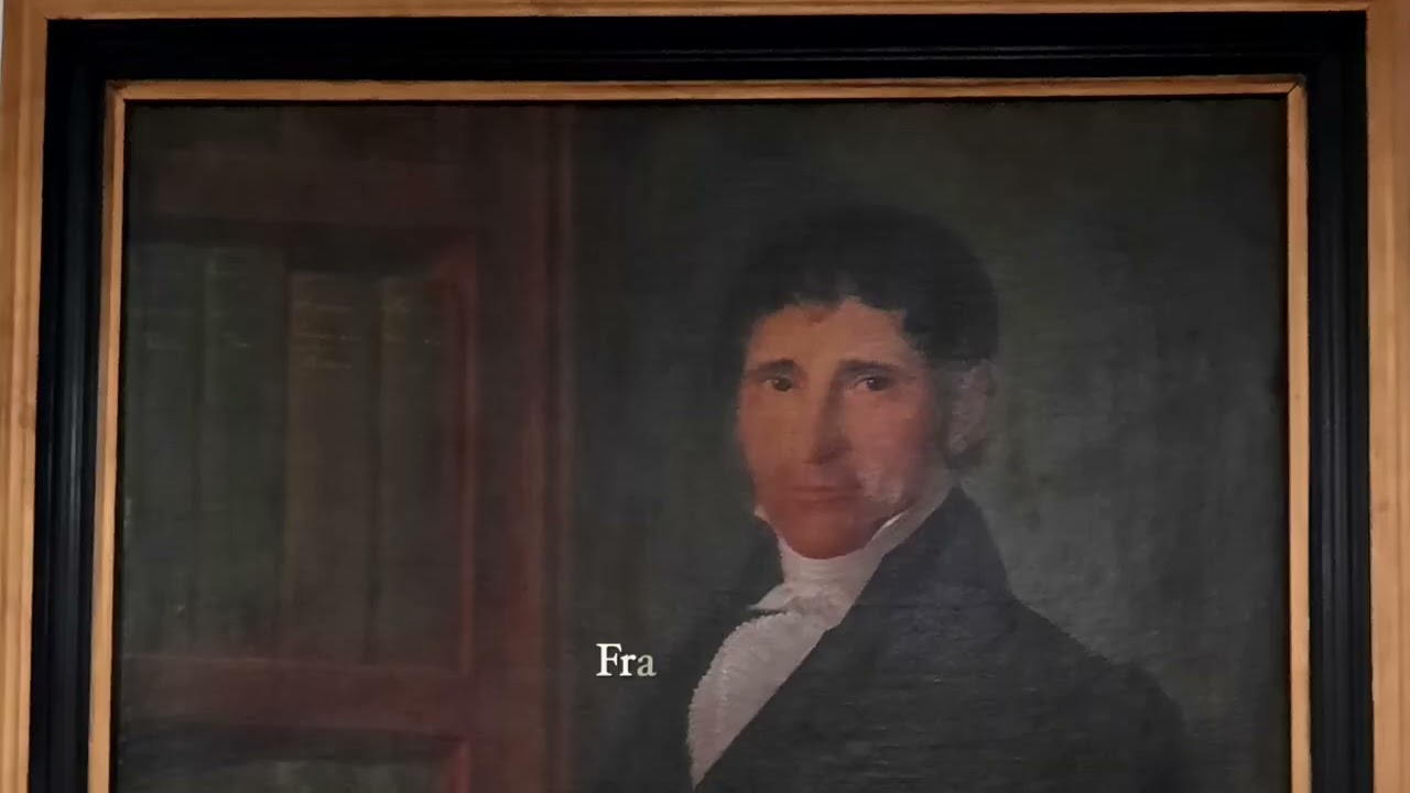
Francesco Loy introduces the anatomical waxes at the University of Cagliari, with close views of the preparations made by Susini and his collaborators.
University of CagliariWatch on YouTube ↗Susini continued to teach drawing as well as model anatomy. He died in Florence in 1814; the museum’s wax workshop survived until 1893. His contribution had often been absorbed into the name of its director, and descriptions of the great collections spoke of Fontana’s waxes. In Cagliari, a more explicit record remained. Every one of the twenty-three cases carried Susini’s signature and date.
Further reading
- Anna Maerker, ‘Turpentine Hides Everything’: workshop organisation and the archival evidence
- Alessandro Riva, Clemente Michelangelo Susini, Dizionario Biografico degli Italiani
- University of Florence, the anatomical wax collection at La Specola
- Marta Poggesi, the anatomical wax collection, in The Collections of La Specola, 2009
- Alessandro Riva and colleagues, The evolution of anatomical illustration and wax modelling in Italy, 2010
- Alessandro Riva and Gabriele Conti, Art and Science in the anatomical waxes of Susini-Boi, 2015
- Roberta Ballestriero, Anatomical models and wax Venuses, 2010
- Josephinum, the wax-model collection and its journey to Vienna
- Ernst Kenndler and Franz Mairinger, chemical analysis of the Josephinum waxes, 1990
- Thomas M. van Gulik, The wax liver model at Josephinum, 2025